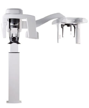
CBCT Technology In Staples Mill
CBCT (Cone Beam Computed Tomography) technology is a form of medical imaging that employs a cone-shaped X-ray beam, a two-dimensional detector, a rotating gantry, and advanced software algorithms to reconstruct detailed 3D images of a patient’s internal structures.

CBCT Technology
What Is CBCT?
Cone Beam Computed Tomography (CBCT) is a specialized medical imaging technique that generates three-dimensional images of internal structures within a patient’s body. It bears resemblance to traditional computed tomography (CT) imaging; however, CBCT utilizes a cone-shaped X-ray beam to capture more detailed images while minimizing radiation exposure. In dentistry, CBCT technology plays a crucial role in diagnosing, treatment planning, and surgical guidance.
By employing a cone-shaped beam, CBCT ensures precise radiation targeting to the specific area of interest, thereby reducing the risk of side effects and exposure to surrounding tissues. The resulting CBCT images provide detailed anatomical information, enabling accurate surgical planning and guidance.
CBCT technology holds immense significance in dentistry for the diagnosis and treatment of various conditions such as tooth decay, jaw disorders, and dental implants. The precise 3D images it produces of the teeth, jaws, and surrounding structures empower dentists to plan and execute complex dental procedures, including orthodontic surgery, root canal treatments, and dental implant placements. Overall, CBCT has revolutionized dental care, providing patients with more accurate and effective treatment options while minimizing risk and discomfort.
How Has CBCT ‘Revolutionized’ Dental Imaging?
The introduction of CBCT technology has brought about a revolutionary transformation in the field of dental imaging. With its advanced capabilities, it enables the production of high-resolution 3D images depicting the teeth, jawbones, and surrounding structures. These intricate images provide dental professionals with the precision needed to accurately diagnose and treat a wide range of dental conditions, ranging from tooth decay to complex surgical procedures.
In contrast to traditional X-rays that often require multiple images to capture different angles of the mouth, CBCT technology significantly reduces radiation exposure for patients. This decrease in radiation-related risks has made the procedure safer and more secure for patients.
Furthermore, the precise images rendered by CBCT technology have resulted in less invasive dental treatments, leading to shorter recovery times and reduced discomfort for patients.
Bid farewell to traditional X-rays and embrace the game-changing high-resolution 3D images of CBCT in dental imaging!
CBCT Benefits
Accurate Diagnosis
CBCT technology enables dental professionals to obtain high-resolution 3D images of teeth, jawbones, and surrounding structures, thereby enhancing the accuracy of dental condition diagnoses compared to traditional X-rays.
Minimized Radiation Exposure
In contrast to traditional X-rays, CBCT technology minimizes radiation exposure for patients, resulting in a reduced risk of radiation-related side effects and enhancing overall safety during the procedure.
Precise Treatment Planning
By utilizing the detailed and precise images produced by CBCT, dental professionals can plan and perform intricate dental procedures, including dental implant placement, with enhanced accuracy and precision.
Less Invasive Procedures
The implementation of CBCT technology has facilitated dental treatments that are less invasive, leading to shorter recovery times and reduced discomfort for patients. Moreover, CBCT has significantly enhanced the overall dental experience, making it more comfortable and stress-free for patients.


